


The unseen world beneath our feet and within a single water droplet has once again taken center stage. Nikon's Small World Photomicrography Competition for 2025 has unveiled its latest collection, showcasing the intricate beauty and surprising diversity of life forms that exist beyond the naked eye. This year's winning images offer a rare glimpse into a universe of minute details, from the delicate structures of a fern to the complex cells within a living organism.
Key Takeaways
- Nikon's Small World Photomicrography Competition highlights microscopic life.
- The 2025 collection features diverse subjects, including plants, insects, and cells.
- Winning images reveal previously unseen details of nature.
- The competition aims to make science accessible through stunning visuals.
Exploring the Microscopic Frontier
For decades, Nikon's Small World competition has invited scientists and photographers to share their unique perspectives of the microscopic realm. Each year, the entries push the boundaries of what can be captured through a microscope, turning scientific observation into high art. The 2025 installment continues this tradition, delivering images that are both scientifically significant and visually captivating.
These photographs do more than just document; they invite viewers to consider the vast complexity present in even the smallest parts of our environment. The competition serves as a powerful reminder that our world holds countless wonders, many of which remain hidden until magnified thousands of times.
Did You Know?
The first Nikon Small World competition was held in 1975, making this year a significant milestone in its history. It has since become one of the most respected photomicrography contests globally.
Diversity of Life Under the Lens
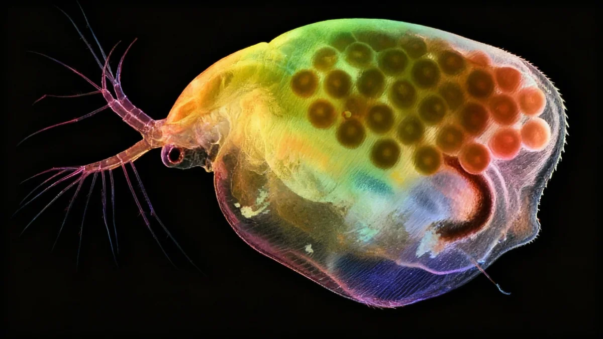
This year's winning entries span an impressive range of subjects, illustrating the breadth of microscopic life. One image depicts a pregnant water flea, known scientifically as Daphnia, captured in stunning detail. This common freshwater organism, often no larger than a grain of sand, reveals intricate internal structures and developing offspring.
Another striking photograph features a rice weevil, Sitophilus oryzae, perched on a single grain of rice. This image highlights the insect's elaborate exoskeleton and its interaction with its food source. Such close-up views provide valuable insights into the behavior and anatomy of these tiny creatures.
"These images bridge the gap between scientific research and artistic expression, making the unseen accessible to everyone," commented one expert. "They spark curiosity and encourage a deeper appreciation for the natural world around us."
Botanical and Cellular Intricacies
The plant kingdom also offered rich subject matter for this year's competition. Images of sunflower trichomes, which are hair-like outgrowths on plants, reveal their delicate and varied forms. These structures play crucial roles in plant defense and water retention, often unseen without magnification.
Another notable entry captured the autofluorescence of lily flower pollen. This technique uses a microscope to make the pollen glow, revealing its unique patterns and textures in vibrant colors. Such images are not only beautiful but also aid in understanding plant reproduction and allergies.
What is Photomicrography?
Photomicrography is the art and science of taking photographs through a microscope. It combines advanced optical technology with photographic skill to capture images of subjects too small to be seen by the naked eye. This field is essential for scientific research, education, and artistic expression.
Unveiling the Inner Workings of Organisms
Beyond whole organisms and plant parts, the competition also delved into the cellular level. One powerful image presented a dedifferentiated liver cell. Dedifferentiation is a process where specialized cells revert to a simpler state, often studied in cancer research and regenerative medicine. This photograph offers a crucial visual aid in understanding cellular biology.
Another fascinating entry showcased colonial algae spheres, specifically Volvox, floating within a single drop of water. These green algae form hollow spherical colonies, with individual cells connected by cytoplasmic strands. Observing them provides insight into early forms of multicellular life and cooperative behavior.
- Key Subjects: Pregnant water fleas, rice weevils, fern spore sacs, sunflower trichomes, lily pollen, dedifferentiated liver cells, colonial algae.
- Techniques Used: Various microscopy techniques, including autofluorescence, to enhance visibility and detail.
- Geographic Diversity: Entries came from photographers across the globe, including China, Germany, Panama, and the United States.
The Art and Science of Small Worlds
The Nikon Small World competition consistently demonstrates the powerful synergy between art and science. Each photograph is a testament to both technical skill and artistic vision. The photographers, often researchers themselves, employ advanced microscopy techniques to illuminate details that would otherwise remain hidden.
These images serve as important tools for education, allowing students and the public to visualize complex biological concepts. They transform abstract scientific ideas into tangible, breathtaking visuals, fostering a greater understanding and appreciation for the intricacies of life.
The full gallery of winning images and honorable mentions is available online, providing a comprehensive tour of these miniature marvels. Each image tells a story, offering a unique window into the vast and diverse universe that exists at the microscopic scale.