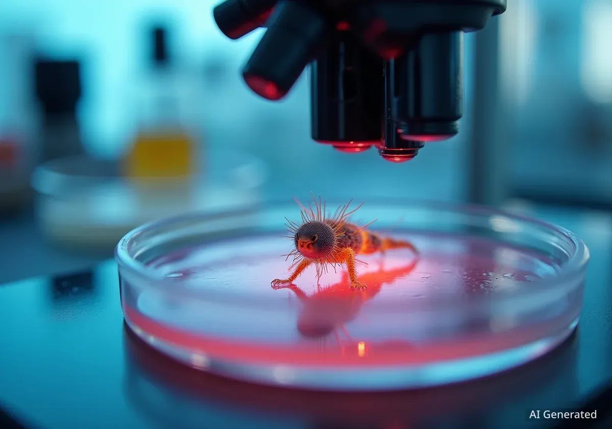
The annual Nikon Small World in Motion competition has announced its 2025 winners, showcasing breathtaking videos of microscopic life and phenomena. These captivating entries reveal intricate details of the natural world that are otherwise invisible to the human eye, ranging from flower self-pollination to the metamorphosis of marine organisms.
The competition highlights the intersection of science and art, bringing together researchers and enthusiasts who use advanced microscopy techniques to capture stunning visuals. This year's entries provide a unique glimpse into biological processes and chemical reactions on a tiny scale, offering both scientific insight and visual appeal.
Key Takeaways
- Jay McClellan won first place for a timelapse of thymeleaf speedwell self-pollination.
- Videos featured diverse subjects like Volvox algae, tardigrades, and cyanobacteria.
- The competition received submissions from scientists and photographers globally.
- Judges selected five top winners and 19 honorable mentions.
- Nikon Small World photo competition winners will be announced on October 15.
Top Honors for Microscopic Flower Pollination
Michigan-based photographer Jay McClellan secured the top prize in the 2025 Nikon Small World in Motion competition. His winning entry was a remarkable timelapse video documenting the self-pollination process of a thymeleaf speedwell flower. McClellan utilized image stacking techniques to achieve a 5x magnification, revealing the delicate movements and structures involved in this biological event.
Winning Technique
Jay McClellan's first-place video employed image stacking. This method combines multiple images taken at different focus points to create one image with a greater depth of field, essential for clear microscopic footage.
In addition to his first-place win, McClellan also received an honorable mention. This second recognition was for a video showing the dissolution and crystallization of cobalt, copper, and sodium chlorides. This entry demonstrated his skill in capturing both biological and chemical processes at a microscopic level.
Diverse Subjects and Advanced Techniques
The Nikon Small World in Motion competition celebrates the diversity of microscopic life and the innovative methods used to document it. Entrants from around the globe submitted videos showcasing a wide array of subjects.
"The competition is a testament to the incredible beauty and complexity of the world beyond our naked eye," a competition spokesperson stated. "It inspires both scientific curiosity and artistic appreciation."
These submissions often employ specialized microscopy, including polarized light, fluorescence, and advanced timelapse photography. The goal is to make the unseen visible, providing new perspectives on fundamental biological and chemical interactions.
About Nikon Small World
The Nikon Small World competition, established in 1975, is a leading forum for showcasing the beauty and complexity of life as seen through the light microscope. The 'In Motion' category specifically focuses on video entries, capturing dynamic processes over time.
Notable Entries from Global Scientists
Scientists and enthusiasts worldwide contributed to the competition, highlighting unique microscopic phenomena. Their work provides valuable insights into various fields, from marine biology to cellular pathology.
- Dr. Alvaro Migotto, a scientist based in São Paolo, Brazil, documented the metamorphosis of a microscopic marine mollusk larva. His video captured the intricate changes as the larva transformed, offering rare footage of this developmental stage.
- Penny Fenton recorded a tardigrade, also known as a 'water bear,' moving across an algae colony. Tardigrades are known for their resilience and unique appearance, making them popular subjects in microscopy.
- Benedikt Pleyer from Germany captured dozens of cyanobacteria filaments using polarized light. This technique enhances visibility of structures that might otherwise be difficult to observe, revealing the organized movement of these microorganisms.
Second and Third Place Winners
The second-place award went to Benedikt Pleyer for his video of Volvox algae. This footage showed the algae swimming within a water drop placed in the central opening of a Japanese 50 Yen coin. This creative presentation combined everyday objects with microscopic wonders.
Dr. Eric Vitriol from the U.S. secured third place. His video focused on actin and mitochondria within mouse brain tumor cells. This entry offered a detailed view of cellular components, which is crucial for understanding disease mechanisms. According to Dr. Vitriol, his work aims to provide clearer images for medical research.
Honorable Mentions and Upcoming Announcements
Beyond the top three, judges recognized 19 additional entries with honorable mentions. These included a video by Wim van Egmond from The Netherlands, showcasing the 'hat thrower fungus' (Pilobolus) on rabbit dung. Janosch Waldkircher from Switzerland also received an honorable mention for an excerpt from a video composed of 7,073 individual images, featuring a male dung beetle (Sulcophanaeus imperator).
Other honorable mentions included:
- Jay McClellan (U.S.) for the dissolution and crystallization of cobalt, copper, and sodium chlorides.
- Dr. Maik C. Bischoff (U.S.) for a developing testis of a fly, showing the actin cytoskeleton (teal) and nuclei (red).
- Dr. Alvaro Migotto (Brazil) for a marine mollusk larva before and after metamorphosis.
- Benedikt Pleyer (Germany) for cyanobacteria (Oscillatoria princeps) filaments from Ishigaki, Japan.
These diverse subjects emphasize the breadth of scientific and artistic exploration within the microscopic world. The full gallery of winning videos is available on the Nikon Small World website. Enthusiasts of microscopic photography should also anticipate the announcement of the Nikon Small World photo competition winners, scheduled for October 15.
The Impact of Micro-Videography
Micro-videography plays a vital role in both scientific research and public education. By capturing dynamic processes, these videos help scientists understand complex biological functions, cellular interactions, and chemical reactions over time. For the general public, they offer an engaging and accessible way to appreciate the hidden beauty and complexity of the natural world.
The competition encourages innovation in microscopy and photographic techniques. It also fosters a global community of researchers and artists who share a passion for exploring the very small. The continuous advancements in imaging technology allow for increasingly detailed and vivid portrayals of microscopic life, pushing the boundaries of what can be observed and understood.


